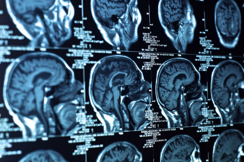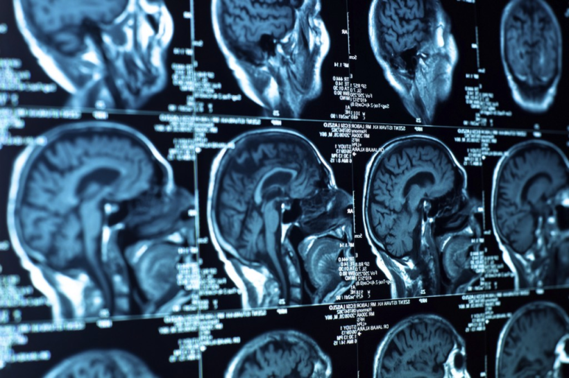
As you may have heard at some point in your life, the human body (on average) is about 60% water (by weight). Water, as you may remember from virtually any chemistry or biology class you’ve ever taken, is a fluid whose molecules have the chemical formula H2O, meaning that each water molecule contains 2 Hydrogen atoms & 1 Oxygen atom. Each of these atoms has a nucleus at its center, which contains positively charged protons & neutral charge neutrons. Under general conditions, the Hydrogen atoms in these water molecules tend to be oriented randomly in your body as they spin about their axes. Once you enter the MRI machine & it boots up, the machine creates a magnetic field which tends to align the spin axes of these Hydrogen atoms with the magnetic field lines (which run along the length of the MRI machine) due to the interaction between the impressed magnetic field & the positively charged protons in the nuclei of the Hydrogen atoms. Most of the Hydrogen atoms either align their axes pointing toward your feet, or towards your head, but there is a small percent that remain unmatched with atoms facing in the opposite direction. When multiplied by the crazy number of Hydrogen atoms in your body, this small percentage becomes quite significant. At this point, the machine begins to emit a Radio Frequency (RF) pulse that excites these unmatched atoms at their resonant frequencies, causing them to align in a specific direction & spin at a certain frequency. When the pulse is discontinued, the unmatched atoms revert to their previous state, releasing energy in the process, & thus emitting a “signal”. This signal is sent to a computer, which uses mathematical formulas (i.e. Fourier Transforms) to translate the signal into a piece of the final image. The greater degree to which the atoms were disturbed by the pulsed energy, the darker those atoms appear in the final image, thus leading to an image of varying contrast. Another major factor in the final appearance of the image is the relaxation rate of the various tissues, or how quickly these atoms revert to their undisturbed states in the various tissues.
By utilizing this technology, medical professionals can help to identify health issues & diseases in their early stages, thus helping to save lives. Pretty cool, right?

